


ShoulderIs chronic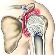 a disease in which articular cartilage tissue is destroyed and thinned, pathological changes in soft tissues occur and bone growths form in the joint area. It is manifested by pain and crunching in the affected area. In the later stages, the range of motion gradually decreases. The pathology is chronic and progressive. The diagnosis is made taking into account the clinical picture and radiological signs. Treatment is usually conservative: physiotherapy, anti-inflammatory drugs, chondroprotectors, exercise therapy. When the joint is destroyed, arthroplasty is performed.
General information
Osteoarthritis of the shoulder is a chronic disease, as a result of the degenerative-dystrophic process, cartilage and other tissues of the joint are gradually destroyed. Usually arthropathy affects people 45 years of age and older, but in some cases (after trauma, inflammation), the disease can develop at a younger age. The disease occurs equally in women and men, and is commonly seen in athletes and those who perform heavy manual labor.
The reasons
The starting point for changes in shoulder osteoarthritis can be both the normal aging of tissues and damage or breakdown of the cartilage structure due to mechanical influences and various pathological processes. Primary arthropathy is usually detected in the elderly, secondary (developing against the background of other diseases) can occur at any age. The main reasons to be considered:
- Developmental disabilities.Pathology can be detected with underdevelopment of the head of the femur or arachnoid space, lipoma of the shoulder and other anomalies of the upper extremities.
- Traumatic injury.Traumatic arthritis usually occurs after a fracture in the joint. One possible cause of the disease may be a dislocation of the shoulder, especially the common joint. Less often, severe bruises act like a traumatic injury.
- Inflammatory processes.The disease can be diagnosed as perennial psoriatic periarthritis, pre-existing nonspecific purulent arthritis and specific arthritis (with tuberculosis, syphilis and some other diseases).
Risk factors
Arthritis is a multifactorial disease. There are a variety of factors that increase your chances of developing this condition:
- Genetic predisposition.Many patients with close relatives also have rheumatic diseases, including other aetiologies (gonarthrosis, coxarthrosis, ankle arthropathy).
- Joint overactivity.It can occur in volleyball players, tennis players, basketball players, sports equipment throwers, as well as in people whose occupation is subject to constant high loads on their hands (hammer, Excavator).
- Other diseases. Arthritis is more commonly found in patients with autoimmune diseases (rheumatoid arthritis), certain endocrine and metabolic diseases, systemic connective tissue insufficiency, and excessive joint mobility.
The likelihood of getting the disease increases sharply with age. Frequent hypothermia and unfavorable environmental conditions have certain negative effects.
Pathogenesis
The main cause of shoulder osteoarthritis is the change in the structure of the articular cartilage. Cartilage loses its smoothness and elasticity, making it difficult to slide joint surfaces during movement. Microtrauma occurs, leading to further deterioration of the condition of the cartilage tissue. Small pieces of cartilage detach from the surface, forming free-standing articular bodies, and at the same time damage the inner surface of the joint.
Over time, the cyst and the synovium thicken, in which areas of capsular degeneration appear. Due to thinning and loss of elasticity, cartilage no longer provides the necessary shock absorption, so the load on the underlying bone increases. Bones deform and grow along the edges. The normal configuration of the joint is disrupted, there are limitations to movement.
Classify
In trauma and orthopedics, a three-stage systematization is often applied, reflecting the severity of the pathological changes and symptoms of shoulder arthritis. This approach allows you to choose the optimal medical tactics, taking into account the severity of the process. The following stages are distinguished:
- The first- no overall change in cartilage tissue. The composition of synovial fluid is changed, the nutrition of cartilage is impaired. Cartilage cannot withstand stress, so joint pain (joint pain) occurs over time.
- Second letter- Cartilage tissue begins to thin, structure changes, surface loses its smoothness, cysts and calcified areas appear deep in cartilage. The underlying bone is slightly deformed, with foci of bony growth along the edges of the joint base. The pain becomes permanent.
- The third day- The cartilage structure is markedly thinned and broken with many areas of destruction. The joint base is deformed. Shows limited range of motion, weakening of the ligamentous apparatus, and atrophy of periarticular muscles.
The symptoms
In the early stages, osteoarthritis patients worry about discomfort or mild pain in the shoulder joint with exertion and some other body parts. Creaking may occur during movement. Joints are not altered externally, no edema. Then the intensity of the pain increases, joint pain becomes a habit, continuously, appearing not only when exercising but also when resting, including at night. Distinctive features of pain syndrome:
- Many patients note the dependence of the pain syndrome on weather conditions.
- Along with soreness, over time, a sharp pain occurs with exertion.
- The pain may be confined to the shoulder joint, radiate down to the elbow joint, or spread throughout the arm. There may be back and neck pain on the affected side.
After a while, the patient began to notice morning stiffness in the joints. The range of motion decreases gradually. After exercise or hypothermia, there may be mild swelling of the soft tissues. With the progression of joint disease, movements become increasingly limited, spastic attacks develop, and limb functions are severely impaired.
Diagnose
The diagnosis is made by an orthopedic surgeon taking into account the characteristic clinical and radiological signs of fibromyalgia of the shoulder. If you suspect secondary arthropathy, consult a surgeon or an endocrinologist. The joint is not changed at first, but later it is sometimes deformed or enlarged. On palpation, the sensation of pain is determined. Restricted movement can be detected. To confirm joint disease, the following are recommended:
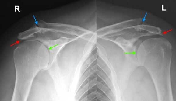
- X-ray of the shoulder joint.Dystrophic changes and marginal growths (osteophytes) are found, in later stages joint space narrowing, deformity and structural changes of the underlying bone are identified. The joint space may be wedge-shaped, with changes in bone tissue and cyst formation visible in the bone.
- Geographical research.In suspected cases, especially in the early stages of the disease, CT of the shoulder joint is performed to obtain more data on the condition of the bones and cartilage. If necessary to assess the condition of the soft tissues, magnetic resonance imaging is performed.
Differential diagnosis
The differential diagnosis of arthropathy is made with gout, psoriasis, rheumatism and reactive arthritis, as well as pyrophosphate arthropathy. With arthritis, blood tests show signs of inflammation; The changes on the radiograph are not obvious, there is no bone formation, there is no sign of deformation of the joint surface.
In psoriatic arthritis, along with joint manifestations, skin rashes are common. In rheumatoid arthritis, a positive rheumatoid factor is identified. With pyrophosphate arthritis and gouty arthritis, biochemical blood tests show corresponding changes (increased uric acid salts, etc. ).
Shoulder joint treatment
The patient is under the supervision of an orthopedic surgeon. It is important to limit the load on the arms, exclude sudden movements, and lift and carry weights for long periods of time. At the same time, it should be noted that inactivity also negatively affects the affected joint. To maintain the muscles in a normal state, as well as restore the shoulder joint, you need to regularly perform therapeutic exercises as recommended by your doctor.
Conservative treatment
One of the most pressing tasks of arthritis is the fight against pain. To eliminate pain and reduce inflammation, the following drugs are prescribed:
- Drugs of general action.NSAIDs are prescribed in tablets during exacerbations. With uncontrolled use, they can irritate the stomach wall, have a negative effect on the state of the liver and metabolism in cartilage tissue, therefore, they should be taken only as prescribed by a doctor. .
- Spot remedies.NSAIDs are commonly used in gel and ointment form. Self-treatment is possible if symptoms arise or worsen. Less commonly, topical hormonal preparations are indicated, which should be applied on the recommendation of a physician.
- Hormones for intra-articular use.In cases of severe pain syndrome that cannot be eliminated by other methods, intra-articular drugs (triamcinolone, hydrocortisone, etc. ) are performed. Blockades are carried out no more than 4 times a year.
To restore and strengthen cartilage in the 1st and 2nd stages of osteoarthritis, agents from the group of chondroprotectors are used - drugs containing hyaluronic acid, chondroitin sulfate and glucosamine. Treatment courses are long (from 6 months to a year or more), the effect is noticeable after 3 months or more.
Physiotherapy treatment
With degenerative shoulder disease, massage exercises, physiotherapy, physiotherapy are actively applied. During remission, the patient is referred to a spa treatment. Application:
- mud and paraffin therapy;
- medicinal leaf bath;
- magnetic therapy and infrared laser therapy;
- supersonic.
Surgery
At stage 3 of the disease, with significant destruction of cartilage, limited mobility and disability, joint replacement is performed. Referrals for surgery are given taking into account the age of the patient, his level of activity, the presence of severe chronic diseases. The use of modern ceramic, plastic and metal endoscopic agents allows you to fully restore the function of the joint. The lifespan of the prosthesis is 15 years or more.
Forecast
Arthritis is a long-term, progressive disease. It cannot be completely cured, however, it can significantly slow down the development of pathological changes in the joints, preserving the ability to work and a high quality of life. To achieve the maximum effect, the patient must take his illness seriously and be willing to follow the doctor's recommendations, even during the period of remission.
Preventive
Preventive measures include reducing household injuries, following workplace safety, eliminating undue load on the shoulder joint when performing professional duties, and playing sports. It is necessary for timely diagnosis and treatment of pathologies that can cause the development of joint changes.